Your retina and vitreous are essential parts of your eye that play a crucial role in clear vision. When these structures are affected by diseases or injuries, timely and advanced treatments are necessary. You can consult our retina specialist, Dr. Rajeev Jain, for all your retinal problems and treatments.
Let’s discuss the retinal problems that might affect your eye health.
Retinal detachment is a serious condition where the retina separates from its supportive layers. It requires immediate attention to prevent permanent vision loss.
The retina, a thin layer of tissue at the back of your eye, is vital for converting light into visual signals. When it detaches, it loses access to the oxygen and nutrients supplied by its underlying layers.
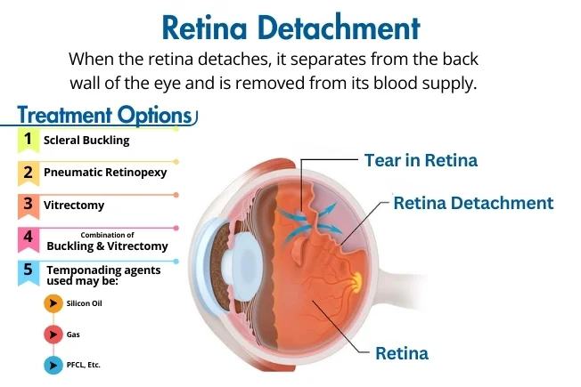
Scleral buckling is a traditional and effective surgery for retinal detachment. It involves:
Best for: Rhegmatogenous retinal detachment (caused by a retinal tear)
Pneumatic Retinopexy is a minimally invasive procedure used for smaller retinal detachments. It involves:
Best for: Simple and upper (superior) retinal tears
Vitrectomy is a highly effective surgery for complex retinal detachments. It involves:
Best for: Complicated cases, giant retinal tears, tractional detachments (due to diabetes), or recurrent detachments.
In some cases, both scleral buckling and vitrectomy are needed for better results.
Best for: Severe or recurrent retinal detachments, cases with multiple retinal breaks.
To keep the retina attached after surgery, the eye is filled with a tamponading agent:
Selection depends on the severity and type of detachment.
Each treatment is chosen based on the type, location, and severity of the retinal detachment. Early diagnosis and surgery are key to preserving vision and preventing blindness. If you notice sudden flashes, floaters, or vision loss, consult an eye specialist immediately.
Know More About Retinal DetachmentThis condition develops in people with diabetes when uncontrolled sugar levels damages the retina’s blood vessels. The severity depends on how long you’ve had diabetes and how well it’s managed.
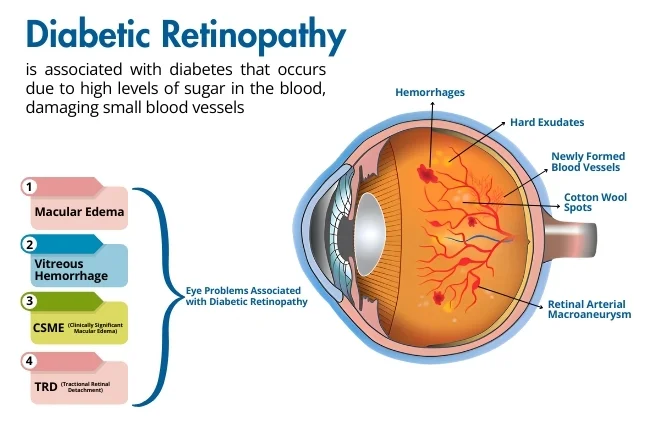
In this early stage, blood vessels may swell and leak fluid. Vision might remain unaffected initially.
This advanced stage involves the growth of abnormal blood vessels that can bleed and cause scarring.
Swelling in the macula (the central part of the retina) leads to blurred vision.
Bleeding into the gel-like vitreous can result in sudden and severe vision loss.
This condition requires urgent treatment due to significant swelling in the macula.
Scar tissue from long-term damage pulls on the retina, causing detachment.
An epiretinal membrane is a thin layer of scar tissue that forms on the retina, causing vision to become blurry or distorted.
The primary treatment is a vitrectomy, where the scar tissue is carefully removed to restore normal vision.
-treatment.webp)
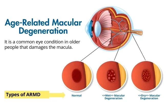
AMD is a common eye condition in older adults that damages the macula, the part of the eye responsible for sharp central vision.
This slower-progressing form is characterized by the build-up of yellow deposits (drusens) in the macula.
This more severe type is caused by abnormal blood vessels leaking fluid or blood into the macula, leading to rapid vision loss.
This advanced form of dry AMD causes significant loss of central vision due to cell degeneration in the macula.
The Amsler Grid is a simple test that helps detect changes in your macula.
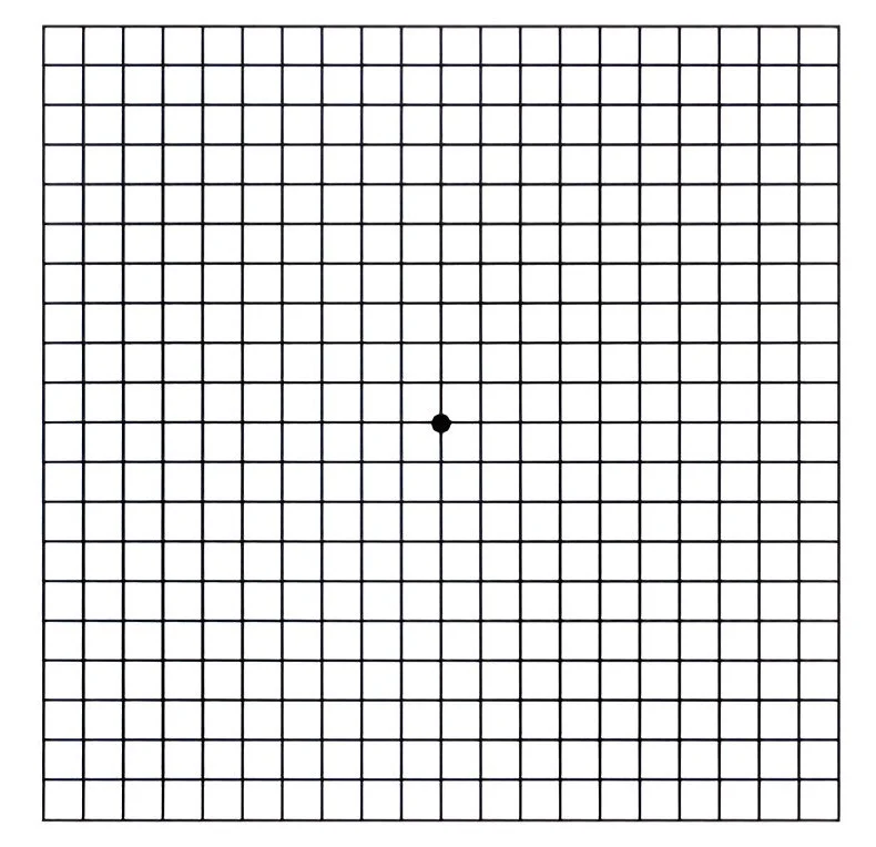
When blood flow is blocked in the retina, it can cause sudden vision loss. Early treatment is crucial to save vision.
Blockage in the main retinal vein causes swelling and bleeding.
Blockage in smaller retinal veins leads to localized damage.
Medications are injected directly into the eye to treat conditions like AMD, diabetic retinopathy, and macular edema.
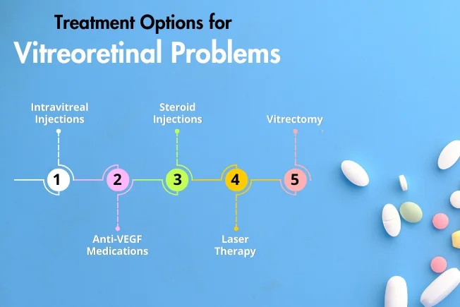
This surgical procedure removes blood, scar tissue, or other debris to restore vision.
If you notice any changes in your vision, such as blurriness, flashes of light, or dark spots, consult our retina surgeon immediately. Early diagnosis and advanced treatment options can preserve your vision and improve your quality of life.
Contact Save Sight Centre today to schedule an appointment and safeguard your vision with the best eye hospital in Delhi
Copyright © 2025 | Save Sight Centre | All Rights Reserved.