Digital Fundus Photography
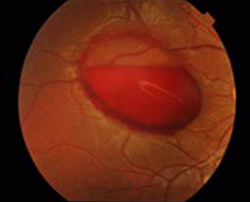
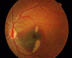
Digital fundus photography (commonly referred to as retinal photography or fundus imaging) plays an integral part in eye health assessment. This non-invasive imaging technique enables detailed retinal examination and early disease diagnosis.
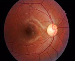
This Fundus eye test involves taking high-resolution images of the retina using a special fundus camera. The retina is light-sensitive tissue at the back of the eye that transmits visual signals to the brain; by taking detailed images of this light-sensitive tissue, digital fundus photography allows eye care professionals to evaluate eye health and detect abnormalities or diseases early.
What are the advantages of Fundus Imaging
This retinal photography offers multiple advantages in eye care:
- Early Detection: This eye test provides high-resolution images of the retina, which enable early detection of various eye diseases, including diabetic retinopathy, glaucoma, and age-related macular degeneration. Early intervention provides greater treatment success.
- Diabetic Retinopathy Monitoring: Diabetic retinopathy is an unfortunate side effect of diabetes that can result in irreparable vision loss if left untreated. Fundus eye test allows regular monitoring of retinal changes among diabetic patients, providing early intervention and management.
- Glaucoma Evaluation: Glaucoma is a progressive eye condition characterized by optic nerve damage. This eye test helps assess and track changes over time within the optic nerve, aiding diagnosis and management of glaucoma.
- Age-Related Macular Degeneration: Age-related macular degeneration (AMD) is one of the primary causes of vision loss among older adults, making digital fundus photography an invaluable way of early diagnosis and monitoring of AMD allowing timely interventions to safeguard vision.
How Does Fundus Photography Work?
It involves several steps for capturing detailed images of the retina:
- Fundus Camera: A fundus camera is a specialized device equipped with high-resolution camera and lenses specifically designed to capture images of the retina, connecting directly to a computer or digital imaging system for image storage and analysis.
- Pupil Dilation: Prior to taking images, patients' pupils are dilated using eye drops in order to provide a better view of the retina and allow for the capture of detailed images. Pupil dilation enables a clearer perspective when viewing detailed retinal images and allows more accurate captures.
- Image Capture Process: Once in position, a patient and fundus camera are aligned for image capture. A flash of light illuminates their retina before multiple images from different angles are captured by the fundus camera.
- Optic Nerve Analysis: Once captured images have been processed and reviewed by eye care professionals such as optometrists or ophthalmologists, they are evaluated in terms of optic nerve, blood vessels, retinal structures and overall eye health to detect any abnormalities or signs of disease.
Preparation for this eye test
To ensure successful digital fundus imaging, certain preparations are necessary:
- Pupil Dilation: Before beginning the procedure, patients' pupils will be dilated using medicated eye drops in order to obtain clear and detailed images of their retina. An eye care professional will give instructions regarding use and any precautions required during dilation.
- Instructions for Patients: Prior to their procedure, patients will often receive specific instructions such as avoiding caffeine or medication that can affect pupil dilation. It is essential to heed such directions in order to achieve accurate results.
Procedure of Fundus Imaging
Digital fundus photography involves three steps.
- Patient Positioning: Once comfortable, either sitting or lying down in front of a fundus camera, an eye care professional ensures that both their head and chin are adequately supported during image capture to minimize movement during this process.
- Image Capture Process: Once aligned, the fundus camera takes multiple images at rapid succession of each eye of a patient. A flash of light illuminates their retina before taking multiple shots quickly in succession.
- Duration of Procedure: The length of a procedure depends on factors like its complexity and patient cooperation; typically however, taking just minutes to capture all necessary images.
Common Eye Conditions Diagnosed with this Eye Exam
This eye exam plays a vital role in diagnosing and monitoring various eye conditions:
- Diabetic Retinopathy: Digital fundus photography provides an effective means of early identification and monitoring diabetic retinopathy, which affects the retina's blood vessels. This technology can assess any blood vessel changes, hemorrhages or fluid accumulation which occurs as a result of diabetes.
- Glaucoma: Digital fundus photography helps diagnose and monitor glaucoma by evaluating optic nerve and retinal structures, including cupping or thinning changes of optic nerve appearances that may indicate disease progression. Images captured can reveal such changes.
- Macular Degeneration: Age-related macular degeneration affects the macula of the retina, located at its central part. Fundus photography helps detect any signs of macular degeneration such as drusen deposits or pigmentary changes, so as to better assess signs of macular degeneration in an earlier stage.
- Retinal Detachment: This eye test can also assist in the detection of retinal detachment, an often serious condition wherein the retina detaches from its supporting tissue and separates from itself. Images help visualizing any tears, breaks or shifts within the retina that might exist as visual clues for diagnoses.
Is digital fundus photography safe?
This is generally safe. It is non-invasive, painless, and usually poses minimal risks or side effects; although using eye drops to dilate pupils may temporarily blur vision and cause light sensitivity; but these side effects usually dissipate quickly.
Can this eye exam detect all eye conditions?
It is an effective imaging technique, but it may not detect all eye conditions. While retina and optic nerve conditions may be easier to spot using digital fundus imaging alone, certain conditions may need additional tests or examinations in order to obtain a comprehensive assessment.
Can digital fundus photography be performed on children?
Yes, It can be carried out safely on children. Modern fundus cameras feature child-friendly features to make use easy for Pediatric patients and ensure comfort during the procedure. Cooperation from children during this procedure should always be prioritized.
Are there any side effects?
Mostly side effects consist of temporary blurry vision and light sensitivity; these tend to resolve on their own in short order. If any unusual symptoms continue or worsen, however, it is wise to consult an eye care professional immediately.