FFA and ICG

What is FFA and ICG Eye Test?

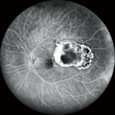
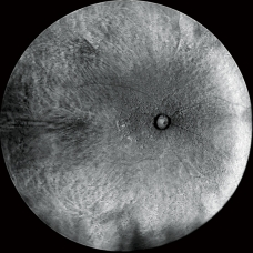
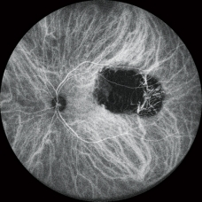
The FFA (Fundus Fluorescein Angiography) and ICG (Indocyanine Green Angiography) eye tests are procedures that help doctors see the blood vessels in the back of your eye. These test help doctors find problems like leaky blood vessels, blockages, or abnormal growths that can affect your vision. These are commonly used to diagnose and monitor conditions like diabetic retinopathy and macular degeneration.

These angiography involve injecting a fluorescein dye into a vein. The dye glows green when exposed to blue light, helping it reach the eye's blood vessels quickly. Doctors then take pictures at different times to see how the dye moves through these vessels. This helps identify abnormal blood flow or leaks in the retina.
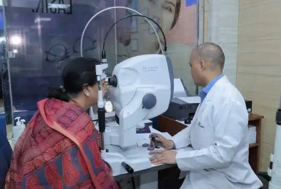
ffa-and-icg-eye-test

Eye Problems Diagnosed by FFA and ICG Eye Tests

The following process is followed commonly:
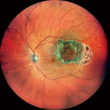
  • Diabetic Retinopathy: Detects abnormal blood vessels and leakage.
  • Age-Related Macular Degeneration (AMD): Identifies blood vessel abnormalities like choroidal neovascularization.
  • Retinal Vein Occlusion: Assesses blockages in retinal veins and associated macular edema.
  • Retinal Artery Occlusion: Diagnoses blockages in retinal arteries.
  • Macular Edema: Detects fluid buildup in the macula.
  • Retinal Detachment: Helps identify the extent and location of detachment.
  • Retinal Tumors: Evaluates the blood supply and nature of retinal tumors.
FFA and ICG Eye Test
  • Choroidal Neovascularization: Found in conditions like AMD, shows new, abnormal blood vessels.
  • Central Serous Chorioretinopathy (CSC): Detects fluid buildup under the retina.
  • Uveitis: Identifies inflammation and blood vessel leakage in the eye.
  • Branch Retinal Vein Occlusion (BRVO): Identifies blockages in smaller retinal veins.
  • Cystoid Macular Edema: Detects cyst-like swelling in the macula.
  • Choroidal Melanoma: Evaluates the blood supply to eye tumors.

Steps Involved in the Test Explained Simply

  • Initial Examination: Your vision is checked and the pressure inside your eyes is measured.
  • Dilation: Eye drops are used to widen your pupils.
  • BP Measurement and Avil Tablet: Your blood pressure is checked, and you are given an Avil tablet to prevent allergic reactions.
  • Injecting Dye: A dye is injected into your arm to help visualize the blood vessels in your eyes.
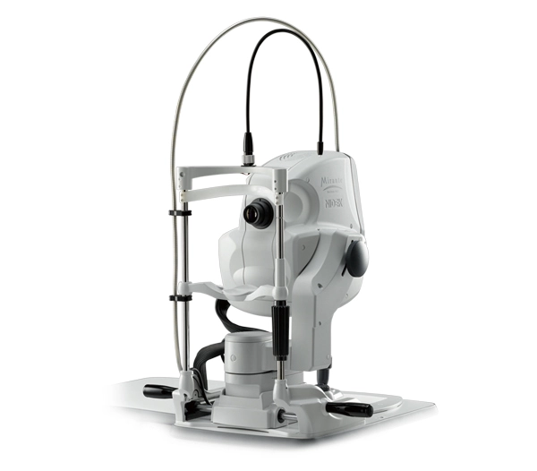
  • Clicking Photographs: A special camera takes pictures of your eyes as the dye moves through the blood vessels.

Benefits of Indocyanine Green Angiography (ICG)

  • Detailed Imaging: Provides clear images of the choroidal blood vessels beneath the retina.
  • Diagnosis of Specific Conditions: Helps diagnose conditions like polypoidal choroidal vasculopathy and central serous chorioretinopathy, choroidal tumors, wet ARMD.
  • Reduced Interference: The dye used in ICG is less likely to interfere with images, allowing for more accurate results.
  • Infrared Light Use: Utilizes infrared light, which penetrates deeper layers of the eye, providing more comprehensive views.
FFA and ICG Eye Test

Benefits of FFA

  • Retinal Visualization: Offers detailed images of the retina's blood vessels, aiding in the diagnosis of retinal conditions.
  • Detection of Abnormalities: Helps detect microaneurysms, hemorrhages, and other abnormalities in diabetic retinopathy and macular degeneration.
  • Treatment Planning: Provides essential information for planning treatments like laser photocoagulation and anti-VEGF injections.
  • Monitoring Progress: Useful for monitoring the progression of retinal diseases and the effectiveness of treatments over time.

Things to Do Before and After FFA and ICG Eye Tests

Before the Test:

  • Inform Your Doctor: Let your doctor know about any allergies, especially to dyes or medications.
  • Eat Light: Have a light meal before the test to prevent nausea.
  • Medication List: Provide a list of your current medications to the healthcare team.
  • Bring Sunglasses: Your eyes will be dilated, so bring sunglasses to protect your eyes from bright light after the test.
  • Arrange Transportation: The dilation will blur your vision, so arrange for someone to drive you home.

After the Test:

  • Avoid Bright Lights: Your eyes will be sensitive due to dilation; wear sunglasses and avoid bright lights.
  • Monitor for Side Effects: Watch for any allergic reactions or side effects like nausea, dizziness, or skin rash.
  • Hydrate Well: Drink plenty of water to help flush the dye out of your system.
  • Follow Up: Attend any follow-up appointments to discuss test results and next steps.
  • Rest: Take it easy for the rest of the day as your vision may be blurry and you might feel a bit tired.

FAQs

FAQs related to Cataract Removal

Que 1: Are there any side effects?

Ans: During the injection you may feel warm or experience a hot flush. This only lasts seconds then disappears. Your skin will be pale yellow and your urine coloured fluorescent green. This is entirely normal and may take two days to wear off.


Que 2: Can I eat and drink before the test?

Ans: Yes. It is advisable to eat a light meal before the test. If you have diabetes you must ensure you have had enough to eat.


Que 3: Can I come alone or drive home?

Ans: No. You are always advised not to come alone for angiography. The drops and bright light from the camera will blur your vision for a short time. You may not be able to drive back home.


Que 4: Should I take my normal medication?

Ans: Yes, all your regular medication should be continued. You will be asked before the test what medication you are taking.


Que 5: Should I inform you of my past medical history?

Ans: Yes, this is very important. Also inform us of any allergies that you may have. If you think that you may be pregnant, please inform the nursing/medical staff.


Que 6: When will I get the test reports?

Ans: You can get the reports on the same day.